PENILE AND PREPUTIAL
PROBLEMS
IN THE BULL
M.S. GILL, D.V.M., M.S.
SURGERY OF THE PENIS
Penile Neoplasia
-
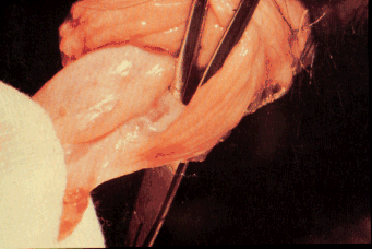
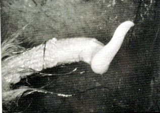
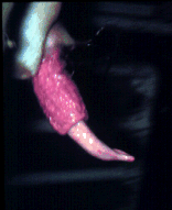
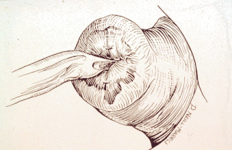
Fibropapilloma of the penis is common and is caused by the
bovine papilloma virus.
-
The virus gains entrance into the skin through
wounds and causes neoplastic growth of fibroblasts.
-
There is no associated
metastasis or local invasion.
-
The condition occurs as a result of
homosexual tendencies of young bulls (1-3 years) housed together.
-
The
warts may be single or multiple and usually occur on the glans and free
portion of the penis.
-
The earliest clinical sign of penile papillomas is
either slight hemorrhage from the preputial cavity following coitus or a
hesitancy or refusal of the bull to breed.
-
Large tumors can lead to either
paraphimosis or phimosis.
-
Removal is achieved with the bull restrained in
a chute or with the use of tranquilization (acepromazine) to facilitate
extension of the penis.
-
Towel forceps in the dorsum of the penis under the
dorsal apical ligament help extend the penis during surgery.
-
The area is
surgically prepared and analgesia is provided by a ring block proximal to
the lesion using 2% lidocaine or
local infiltration around the dorsal nerves (5-10cc of 2% lidocaine)
across the dorsum of the penis near the preputial orifice.
-
A gauze
tourniquet can be placed proximal to the surgery site to provide some
hemostasis.
-
For warts that occur close to the urethral groove, passage of
a urinary catheter is helpful prior to surgery.
-
The papillomas are removed
by dissecting through the epithelium at the base of the growth.
-
Small
vessels are ligated and the epithelium is closed with #2-0 absorbable
suture material.
-
Sexual rest is provided for 2-4 weeks after surgery.
-
Recurrence is common if the bull is in an active stage of the disease.
-
Administration of a wart vaccine (commercial or autogenous) may reduce
recurrence.
Persistent frenulum
-
This condition is characterized by a fibrous band attaching
the prepuce to the free portion of the penis along the median raphe
immediately caudal to the glans.
-
It manifests as a marked ventral
deviation of the bull's penis during attempts at coitus.
-
It is noted
chiefly in Angus and Shorthorn bulls (also reported in Brangus, Santa
Gertrudis, Beefmaster, and Hereford).
-
This condition prevents intromission
in Bos taurus breeds
but may not interfere with breeding in Bos
indicus bulls due to the length of the prepuce.
-
The anomaly is congenital and suspected to be a heritable condition.
- The
epithelial surfaces of the penis and prepuce of bulls are fused at birth.
-
At approximately 4 months, the penis and prepuce begin to separate under
the influence of male hormones.
- Separation should be complete by 8 to 11
months of age.
- The frenulum normally ruptures during separation of the
penis from the prepuce.
- The condition can be easily surgically corrected
but owners should be advised of the possibility that this is a heritable
condition.
- To surgically correct this condition the bull is restrained in
a chute and the penis is extended.
- The frenulum is infiltrated with 5 cc
of 2% lidocaine at the penile and preputial attachments.
- It is then
ligated at each end with #2-0 absorbable suture material and excised.
-
Sexual rest is allowed for 2 weeks.
Penile deviations
-
1. types of deviations
-
- the majority of deviations occur
in the polled beef breeds - polled Hereford and Angus
-
a. spiral or corkscrew - the most common deviation observed
- occurs in bulls between 2 ½ and 5 years of age.
-
Spiral deviation occurs
at the peak of erection.
-
The spiral configuration is caused by the dorsal
apical ligament slipping off to the left hand side of the penis resulting
in a counter clockwise spiral as viewed from the rear.
-
Up to 50% of normal
bulls have been shown to develop spiral deviation during copulation.
-
The
condition is often noticed in normal bulls during masturbation, following
intromission and when erection is produced by an electroejaculator.
-
It is
not considered pathologic until its occurrence is observed on repeated
natural breeding trials in which it occurs prior to entrance into the
vulva and thereby prevents intromission.
-
b. ventral or rainbow deviation -
-
less common than spiral -
-
ventral deviation prevents intromission -
-
this condition can be diagnosed
by electroejaculation -
-
occurs when the ligament is thin and stretched to
the point that it is incapable of holding up the distal portion of the
penis during erection.
-
c. S-shaped deviation
-
- relatively rare -
-
usually occurs in
older bulls with an excessively long penis.
-
The apical ligament is
sufficient in strength but insufficient in length, and the S-shape results
during erection -
-
no surgical techniques have been described for this
deviation.
d. lateral deviations - may occur secondary to trauma of
the penis or prepuce - scars or adhesions of the elastic tissue pulls the
penis to one side or the other.
-
2. fascia lata implant technique - used for repair of both
spiral and ventral penile deviation -
-
the objective of the technique is to
create a firm union of the dorsal apical ligament to the tunica albuginea
to prevent its slippage.
-
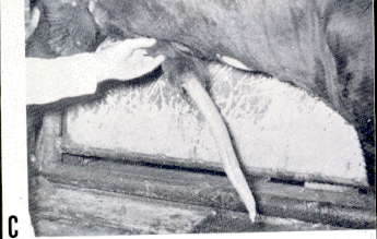
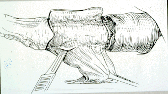
Surgical technique:
-
Withhold food and water for
48 hours,
-
general anesthesia,
-
right lateral recumbency,
-
the preputial
hairs are clipped,
-
penis extended manually, and
-
a towel clamp is placed 5
cm from the apex of the penis under the dorsal apical ligament (avoid
penetration of the urethra and glans penis).
-
The penis is scrubbed and
prepared for surgery then allowed to retract into normal position while
the fascia lata is harvested and prepared.
-
A site over the left lateral
thigh from the patella to the tuber coxae is surgically prepared (20 X 40
cm).
-
A skin incision is made starting 10 cm proximal to the cranial
lateral aspect of the patella and extending 20 cm toward a point halfway
between the tuber coxae and greater trochanter.
-
The incision is extended
through the superficial fascia until the thicker deep layer of fascia lata
is reached.
-
A 3 X 15 cm section of fascia lata is removed over the vastus
lateralis m. and placed in warm saline.
-
The fascia is closed with #2
vicryl and the skin is closed with 0.6 mm Vetafil (caprolactam).
-
The strip
of fascia lata is prepared by removing all loose connective tissue and fat
from both sides and it is then returned to the warm saline solution.
-
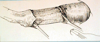
The penis is again extended and held in that position for
the remainder of the surgical procedure.
-
A 15-18 cm incision is made on
the absolute dorsum of the penis (1800
opposite the urethral groove).
-
This incision should be centered at the
preputial reflection.
-
The incision is extended, through the loose fascial
layers in the free portion of the penis and the thin elastic layers in the
preputial portion, to expose the apical ligament.
-
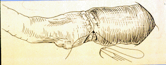
The apical ligament is
incised throughout its length along the dorsum of the penis.
-
This exposes
the tunica albuginea of the penis.
-
The ligament is reflected on both sides
to create a bed for the fascia lata and to remove the thin fascia
separating it from the tunica albuginea.
-
Care should be taken to avoid
damage to the terminal branches of the dorsal vessel and nerve on the
right side - these vessels must not be covered by implant.
-
The fascia lata
implant is placed between the apical ligament and tunica albuginea
beginning as far proximally as possible.
-
Four simple interrupted #0 vicryl
sutures are placed through the fascia lata and into the tunica albuginea
at the proximal aspect.
-
Interrupted sutures are placed along the lateral
margin of the implant at 2-cm intervals, stretching the fascia lata in
both directions.
-
The implant is then sutured at the distal end under mild
tension with three interrupted sutures.
-
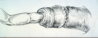
The apical ligament is sutured (#0
vicryl) over the fascia lata graft in a continuous pattern including the
fascia lata in every second or third suture.
-
The elastic layers are closed
in the preputial area with 3-0 vicryl and the epithelium of the prepuce
and free portion of the penis are closed with simple interrupted 2-0
vicryl.
-
The penis is allowed to retract into the preputial cavity.
-
Postop
antibiotics are administered for 5 days.
-
The penis is manually extended
and sutures removed at 10 days.
-
The fascia lata implant becomes homogenous
with both the dorsal apical ligament and the tunica albuginea as early as
30 days post-surgically and is complete by 60 days.
-
These bulls can be
used for breeding at 60 days post-op.
-
The prognosis for return to breeding
soundness is considered more favorable with spiral than ventral
deviations.
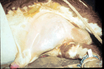
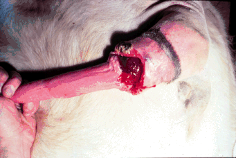
Hematoma of the penis
-
Hematoma of the penis is also referred to as "ruptured
penis", "broken penis" or "fractured penis" -
-
common in bovine species and rare in others -
-
occurs during coitus when
the cow slips or goes down under the weight of the bull or when the penis
is thrust against the escutcheon of the cow during breeding attempts.
-
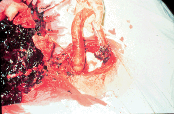
The
corpus cavernosum penis (CCP) is a closed system and during erection,
blood pressures within the CCP may exceed 14,000 mm of Hg.
-
Sudden
angulation of the penis may increase the blood pressure and result in a
hematoma.
-
Penile hematoma results from a tear of the tunica albuginea into
the CCP.
-
The tear usually occurs on the dorsum of the penis at the distal
sigmoid flexure opposite the insertion of the retractor penis muscles; the
tunica albuginea is thinner in this area.
-
The tear is usually 2-7.5 cm
long and transverse - these tears usually do not extend over 180E
circumferentially.
-
The swelling due to hematoma is a result of blood from
the CCP being forced into the peripenile tissues.
-
The owner frequently
first notices the presence of a prolapsed prepuce which may result
secondary to the swelling.
-
Diagnosis is based on physical exam and the
presence of a large swelling immediately cranial to the scrotum.
-
The
swelling is soft until the clot begins to form by about the fourth day.
-
By
10 days the clot begins to organize and the swelling becomes quite firm.
-
Bruising of the skin over the hematoma may be apparent.
-
Size of the
hematoma is not related to the length of the tear in the tunica albuginea
but to the number of coital attempts the bull makes following rupture.
-
Treatment:
-
1. Medical - spontaneous recovery occurs in >50% of the
cases given 90 days sexual rest -
-
therapy includes parenteral antibiotics
(1 week),
-
warm local hydrotherapy (2-3 weeks), and
-
sexual rest for 60-90
days.
-
2. Surgical -
-
should be performed between 5 and 10 days
post-trauma,
-
surgical repair probably reduces the incidence of
complications.
-
A second chance for surgical repair is after 21 days when
fibrosis dissipates.
-
Surgical technique -
-
fast 48-72 hrs. prior to surgery,
-
right lateral recumbency,
-
general anesthesia or heavy sedation/local
analgesia,
-
surgical prep of area.
-
A 20 cm skin incision is made over the
swelling cranial and parallel to the rudimentary teats.
-
The incision is
extended through the subcutaneous tissues until the hematoma is
encountered dorsal to the penis.
-
The hematoma is incised and the clot
manually removed.
-
The remaining cavity is flushed with dilute Betadine in
sterile saline.
-
The penis and surrounding elastic tissue are exteriorized
through the skin incision.
-
The retractor penis muscles and urethral groove
should be located to aid orientation.
-
The elastic layers are incised
longitudinally on the left lateral aspect of the penis to permit
exteriorization of the tunica albuginea.
-
The rent is debrided and then
apposed with #1 vicryl in a bootlace pattern which is a widely spaced
simple continuous reversed upon itself.
-
The elastic layer is apposed over
the penis and sutured with 3-0 vicryl, simple continuous pattern.
-
The
penis is returned to normal position and the cavity flushed again with
dilute Betadine in saline and dried with sterile towels.
-
The subcutaneous
layers are closed using a continuous pattern with #0 vicryl, the skin is
closed with 0.6 mm vetafil.
-
Postop antibiotics are given for 5 days.
Seroma formation will occur and
should subside about 10 days after the surgery.
- Skin sutures are removed
10 days post-op.
- Sexual rest is provided for 60 days.
-
Undesirable sequella:
-
1. recurrence of the hematoma - common, but
surgical repair lessens the risk - 50% recur with medical treatment, 25%
recur with surgical treatment.
-
2. adhesions of elastic layers to the
tunica albuginea or to the skin preventing complete extension of the
penis.
-
3. analgesia of the penis due to damage of
the dorsal penile nerve may occur
-
a. at time of hematoma.
-
b. several months later due to fibrous
tissue impingement.
-
c. or, when breeding resumes and nerves are
stretched.
-
4. abscessation
-
a. hematogenous route.
-
b. break in aseptic technique during
surgery, abscessation may then lead to adhesions or septic cavernitis.
-
5. vascular shunts
-
- between CCP and dorsal
vessels -
-
no longer a closed system in the CCP---> erection failure.
SURGERY OF THE PREPUCE
-
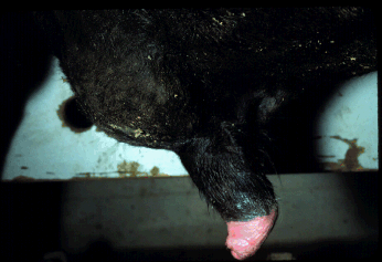
Preputial trauma/laceration/prolapse
-
Habitual prolapse of the prepuce is common
especially in the following breeds:
-
Brahman,
-
Brahman cross,
-
Angus, and
-
polled Hereford.
-
The predisposition to preputial prolapse probably
involves four anatomic features:
-
1. pendulousness of the sheath (prepuce
below the carpus, low preputial angle)
-
2. length of the prepuce
-
3. size of the preputial orifice
-
4. presence of retractor prepuce muscles
-
(frequently absent in polled breeds -
- 1/3 of all polled bulls lack these
small paired muscles)
-
The sheath and prepuce of Bos
indicus bulls are more pendulous
averaging 5.5 cm longer than Bos
taurus breeds.
-
Preputial prolapse and trauma often occurs
one of two ways.
-
Bulls which frequently have some prepuce exposed may
develop abrasions and lacerations of the prepuce from exposure to
environmental factors (including frostbite).
-
This may lead to edema,
further prolapse, more trauma, abscessation and eventually fibrosis of the
preputial tissue.
-
Or, a laceration of the prepuce during breeding may
occur which usually leads to preputial prolapse.
-
Lacerations of this type
occur when there is a "bunching" of excess preputial tissue
immediately prior to the breeding lunge.
-
The tissue usually bursts
ventrally in a longitudinal direction when the prepuce impacts upon the
pubis of the cow.
-
Because of the elastic layers and retraction of the
penis there is a marked tendency for healing to occur in a transverse
manner which shortens the effective length of the prepuce as it heals.
-
The
traumatic injury results in edema formation and preputial prolapse.
-
Treatment is initially aimed at medical management.
-
The prepuce should be
soaked in a warm dilute betadine/epsom salt solution.
-
Coat the prepuce
with "petermycin" (lanolin + scarlet oil + tetracycline).
-
If the
prepuce can be replaced, a sterile drain tube (3/4-1" x 5-6") is
placed in the preputial cavity to hold the prepuce in place.
-
The tube is
taped in place with elasticon taping to the haired sheath only.
-
The wrap
should be changed daily, initially, then as needed.
-
If the prepuce can not
be replaced at first it can be treated as above and the exposed portion
covered with a stockinette.
-
A drain tube is inserted in the preputial
cavity and must extend above the proximal part of the wrap to allow for
urine egress.
-
The prepuce (covered by stockinette) is wrapped with
elasticon applying even pressure.
-
This pressure wrap will decrease the
edema and swelling usually making the prolapse reducible in 1-2 days.
-
Change wrap daily, then as needed.
-
The object of medical treatment is to
get control of edema, cellulitis and necrosis and establish a healthy bed
of granulation tissue.
-
Surgery is indicated if fibrous tissue
development prevents normal extension of the prepuce or if there is
habitual prolapse of the prepuce.
-
If the value of the bull does not
warrant surgery, salvage is an option.
-
The most commonly performed
surgical technique is the reefing procedure (resection-anastomosis).
-
This
technique is employed to revise damaged preputial tissues.
-
General
anesthesia, or tranquilization with a pudental nerve block or ring block
may be used.
-
A penrose drain tourniquet is tied near the preputial orifice
and towel clamps are placed under the dorsal apical ligament to allow
extension of the penis and prepuce during surgery.
-
The site is surgically
prepared and aseptic technique is utilized.
-
Two circumferential incisions
are made a sufficient distance apart to excise the affected tissues.
-
The
prepuce is measured prior to incision to assure that the final preputial
length will be at least twice (2X) the length of the free portion of the
penis.
-
The circumferential incisions are connected by a longitudinal
incision and the affected portion of prepuce is sharply dissected from the
underlying elastic tissues.
-
Dissection should be as superficial as
possible but as deep as necessary in the areas of scar tissue formation.
-
Hemostasis is important and after completion of dissection the tourniquet
is removed and every effort is made to control hemorrhage to prevent
subsequent hematoma/seroma formation.
-
Closure is in two layers, elastic
tissue and skin.
-
An interrupted suture pattern using 2-0 vicryl is used
for both layers.
-
A penrose drain is sutured over the free end of the penis
to prevent urine contamination during healing.
-
The penis is allowed to
return to the preputial cavity and a sterile soft rubber tube (with the
penrose threaded through its lumen) is bandaged in place to retain the
prepuce during healing.
-
The bandage is removed in 5 days.
-
At 10-14 days
the penis is extended and any remaining sutures removed.
-
Sexual rest is
recommended for at least 60 days.
Preputial avulsion
-
Preputial avulsion usually occurs during
semen collection with an artificial vagina (AV).
-
If the AV is too tight, a
transverse or oblique laceration may occur at the dorsum of the preputial
ring or fornix.
-
These injuries should be sutured immediately using 2-0
vicryl in an interrupted pattern.
-
Allow 60-90 days of sexual rest.
Retropreputial abscess
-
This is a frequent sequella to preputial
laceration in Bos taurus
bulls.
-
The bull retracts the prepuce into the preputial cavity after
injury.
-
Drainage is impaired causing abscess formation and the abscess
dissects into the elastic layers.
-
Prognosis is guarded; there is less than
30% chance of return to service.
-
Conservative approaches include immediate
salvage or 6 months pasture rest and re-examination; abscesses will
occasionally effectively heal on their own.
-
More aggressive treatment is
drainage of the abscess into the preputial cavity followed by antiseptic
flushes.
-
If the abscess is drained through the skin, adhesions almost
always develop which will eventually prevent penile extension.
-
Systemic
antibiotics are not that useful once the abscess is formed but may be of
help if cellulitis is present.
- Actinomyces
pyogenes is most frequently
isolated from such abscesses, therefore, penicillin is the antibiotic of
choice.
- Regardless of the method of treatment, possible complications
include stricture formation and adhesion formation resulting in a useless
breeding animal.
|
 Male
Index
Male
Index