Equine
Reproductive
Anatomy
 43-69 43-69

Embryology
- see Pathways to Pregnancy and Parturition
Brain
- You should also review the bovine notes
and Pathways to Pregnancy and Parturition.
- The pineal gland is between the cerebral
hemispheres and is connected by a stalk to the brain.
- The pineal gland secretes melatonin. Melatonin is secreted in
higher amounts in times of increasing darkness, so less melatonin
is released during the cycling season than occurs during the times
of longer daylight.
- Melatonin appears to inhibit cyclicity by
decreasing the amount of GnRH released.
- Some mares with high melatonin, however,
continue to cycle. This may be due to an insensitivity to melatonin
or some other factors may be involved.
Ovaries
- The ovaries are kidney bean shaped and their
size varies with season.
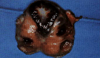
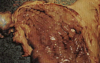
An ovary cut in two showing the hilus at the bottom.
- The ovaries are suspended by the mesovarium
portion of the broad ligament.
- There are three surfaces used to identify
structures on the ovary .
- The cranial pole is attached to the
fimbria,
- The caudal pole is attached to the uterus by the proper
ligament of the ovary
- The lateral and medial surfaces.
- The
dorsal (attached surface) is mesenteric and the ventral (free
surface) is anti-mesenteric.
- The ovulation fossa is at the 'hilus' of the
ovary.
- The entire ovary is covered by peritoneum,
which means that some mares will show pain at ovulation because of
the stretching of the peritoneum.
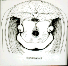
Location
- The ovarian location is somewhat variable, but
is usually at 10 and 2 O'clock positions by the shaft of ileum. They
are always at the end of the uterus, however.
- The ovaries may be behind the broad ligament
and may need to be 'flipped out' from behind the ileum in order to
thoroughly palpate them.
Structure
- The germinal epithelium is on the inside,
rather the outside of the ovary (inside out compared to a cow). This
prevents external ovulation of the oocyte and ovulation is only through
the ovulation fossa at the hilus of the ovary. This internal
location of the germinal epithelium prevents the CL protrusion and
palpation as in the cow.
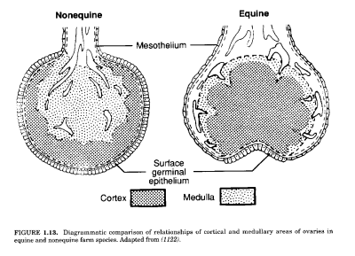
- Only follicles on the equine ovary are readily
palpable.
- The CL cannot be palpated.
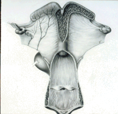
Oviducts
- The oviduct has three main segments, the
fimbria (infundibulum), ampulla and isthmus.
- The fimbria catches the ova for transport to
the remaining segments of the oviduct. There may be cysts present by
the oviducts, which are remnants of the Müllerian (tubo-ovarian
cysts) or Wolffian (epoophoron or paroopheron cysts) ducts. These
cysts are usually not clinically significant, but occasionally may
be confusing during palpation or large enough to physically
interfere with ovulation.
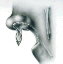
- The uterotubal junction
(UTJ) acts as a mechanical
barrier as is identified as the ovulation papilla in the uterus.

Endoscopic view of the UTJ in a mare.
Uterus
- The uterus is 'T' shaped and suspended by the
mesometrial aspect of the broad ligament. It is usually half
abdominal and half pelvic.
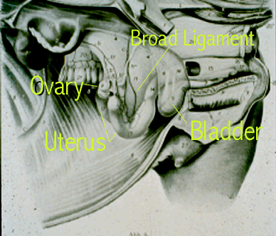
- It is dorsal to the bladder and anterior to the
pelvic brim. The location may vary due to intestinal displacement,
however.
- The broad ligament attaches dorsally to the
uterus (the ventral uterus is free) and then it attaches dorsally to
the sublumbar region from the 3rd-4th lumbar to the 4th sacral
vertebra. The broad ligament may contain large amounts of fat. The
long attachment allows exteriorization surgically.
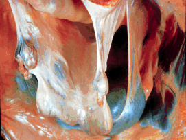
- The endometrial lining consists of hundreds of
folds.
Folds in the endometrium.
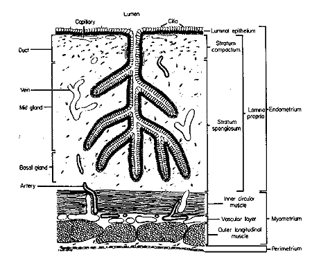 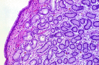
- The knowledge of endometrial histology is
important for assessing fertility.
- The layers include:
- The
epithelium,
- The stratum compactum which lies under the epithelium
and goes down to the glands
- The stratum spongiosum which
includes the glands and goes down to the myometrium.
Cervix
- The cervix in the mare has an internal os and
an external os.
- It has no cartilage and the tone changes during
cycle.
- There are longitudinal folds, of which the
exterior outgrowth is the frenulum. This is seen in the anterior
vagina as closed flower bud in diestrus and a 'wilted rose' during
estrus.
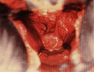 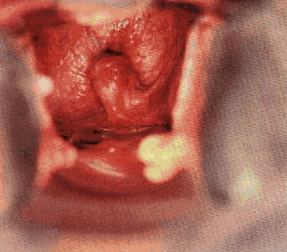
A soft cervix at the left and a tight cervix on the right as seen
during vaginoscopy.
- The cervix forms the final barrier to uterine
contamination.
Vagina
- The vagina is a potential space that is
normally dorsally-ventrally flattened.
- Loss of support by ligaments and poor condition
may cause anterior-ventral sloping of the pelvic floor and allow
urine to pool by the cervical os.
- The vagina ends at the vaginal vestibular
junction.
- Vaginal-vestibular fold
- The vaginal-vestbular fold is the second
barrier to contamination of the uterus.
- It is formed by the end of Müllerian duct.
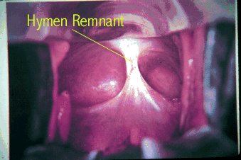
- This is where you may see a persistent hymen.
- The urethra exits posterior to the transverse
fold.
Vestibule
- The vestibule extends from the vagina to the
labia.
- It is of ectoderm (cloacal) origin.
- This is an area for potential tears at foaling,
which are classified as first, second, or third degree tears.Vulvar labia
- The
labia present the first line of defense to contamination of the
reproductive tract.
- They are composed of skin and mucus membranes.
- The desirable conformation is vertical and in
the same plane as the rectum. At least 80 % of the lips should be
below the pelvic brim.
- Poor conformation leads to a weak seal and
allows contamination of the vestibule and vagina.
- Tears, a sunken rectum, the vulva above the
pelvis, and tilted labia all contribute to contamination.
- The causes for poor conformation include tears,
age, and weight loss.
- The 'wind sucker' tests for air entry to the
vagina when the vulvar lips are pulled apart.
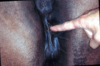
Clitoris/clitoral
fossa
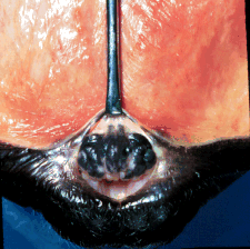
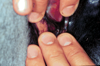
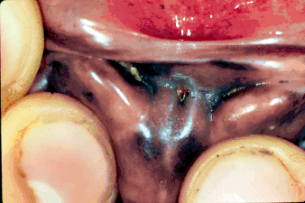
Examination of the clitoris and clitoral fossa.
- The clitoris is inside the lower vulva.
- The clitoris is homologous to the penis and can
be seen everted as 'winking' during estrual teasing. This is because
of the erectile tissue it contains.
- The fossa may harbor contaminants, which
include CEM (Contagious Equine Metritis) and other bacteria.
- In the fossa are the clitoral sinuses, which
are the preferred site to sample for CEM.
Bladder
- The bladder is ventral to the uterus and great
care should be taken not to confuse it with the uterus during
examination.
- The lateral ligament of bladder is posterior to
the broad ligament and contains the round ligament of the bladder
(which is umbilical in origin).
- The ventral ligament of the bladder is from the
urachus.
|
 Equine
Index
Equine
Index